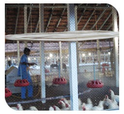
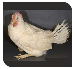
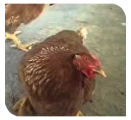
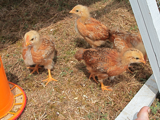
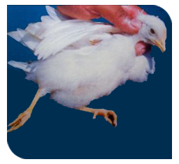
Newcastle Disease (ND) / Ranikhet Disease (RD)
Nature of disease
 |
 |
 |
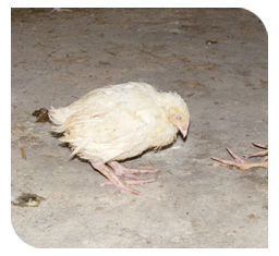
| Depression |
Diarrhoea |
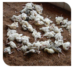
Mortality |
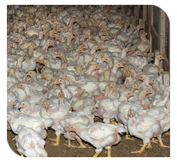
- This is an acute viral disease of poultry characterized by involvement of respiratory system, drop in egg production and mortality as high as 100% in severe cases.
- This virus has zoonotic effect and can causes human deaths.
Causes
|
|
|
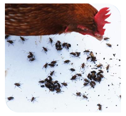
Contamination of water |
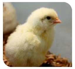
Infected chicken |
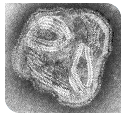
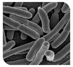
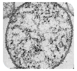
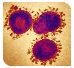
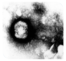
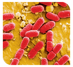
Paramyxovirus |
- Paramyxovirus type1 (PMV-1 belongs to the genus Avulavirus, family Paramyxoviridae).
- Based on the disease produced in chickens, NDVs have been classified into five pathotypes, namely Viscerotropic velogenic (most pathogenic), Neurotropic velogenic, Mesogenic (moderate pathogenic), Lentogenic9 low pathogenic) and Asymptamatic.
- Various physical (heat, irradiation and pH effects) and chemical compounds (potassium permanganate, formalin, ethanol, etc.) could destroy the virus.
- Fumigation of buildings and incubators can be used to kill the virus.
- Infected chickens are the primary source of virus.
- Virus is shed during incubation, during the clinical stage, and for a varying but limited period during convalescence.
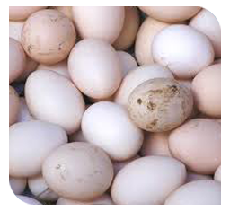
- Virus may also be present in eggs laid during clinical disease and in all parts of the carcass during acute virulent infections.
- Infected birds shed virus in exhaled air, respiratory discharges, and feces, etc. which contaminate feed and water.
- Infection in birds occurs through inhalation and ingestion of contaminated materials.
- Wild and pet birds, movement of people and poultry equipment and even poultry products could aid in spread of infection.
- The virus has been found to survive for several days on the mucous membrane of the human respiratory tract and has been isolated from sputum.
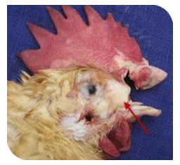
Clinical symptoms
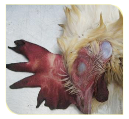
- Twisting of neck and paralysis of wings and legs
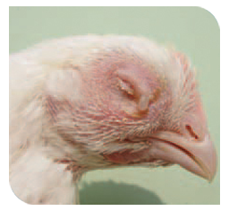
- Cyanosis of comb
- Facial edema
- Diarrhoea
- Drop in egg production
- Sudden death
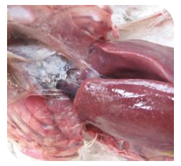
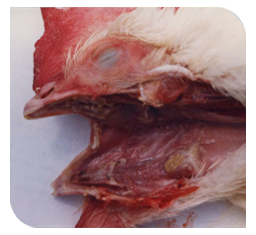
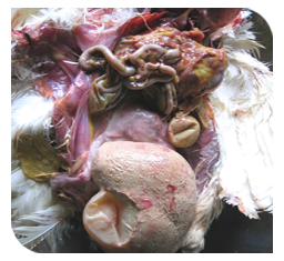
Gross lesions
- Haemorrhage in intestine
- Petechial haemorrhage in proventiculus
- Congestion and mucoid exudates seen in the respiratory tract, especially in trachea.
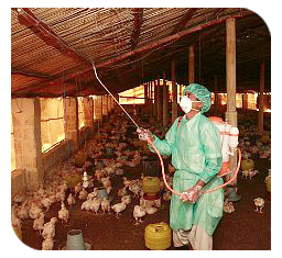
Prevention and control
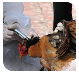
- Disease can be prevented effectively by an integrated approach of vaccination, proper management and strict biosecurity.
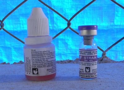
- Live virus vaccines both from lentogenic (La Sota, F, B1) and mesogenic (H, R2B, Mukteshwar) strains are used for induction of good immune response.
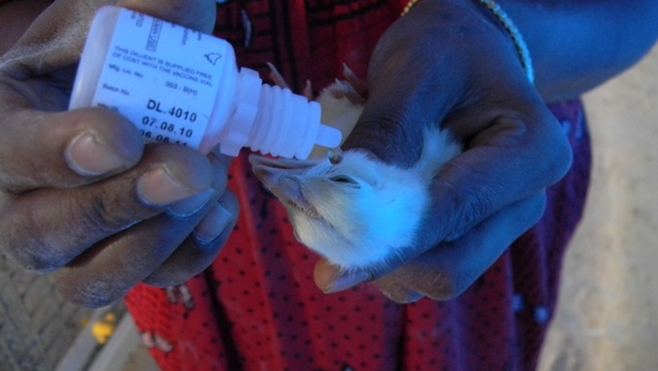
- Healthy chicks are vaccinated as early as day 1-4 of life.
- Lentogenic strains are administered through ocular (eye) or nasal (nostril) route for primary vaccination of birds.
- Mesogenic strains are administered through subcutaneous or intramuscular route at 6-8 weeks of age as secondary vaccine for better protection of longer duration.
- Killed vaccine with oil adjuvant are used in endemic areas and administered intramuscularly/ subcutaneously to maintain high and prolonged antibody titre in layers and breeders and better maternal antibodies in chicks.
- Recommended vaccination schedule for layers:
Age in days |
Name of the vaccine |
Route |
5 |
F/B |
I/o (or) I/n |
27 |
LaSota |
water |
52 |
LaSota |
Water |
64 |
R2B |
I/m |
|
|
|
112 |
LaSota |
water |
280 |
LaSota |
water |
- Regular disinfection of farm premises and equipment with potassium permanganate (1: 1000), sodium hydroxide (2%) or Lysol (1: 5,000) are useful in preventing this disease.
TOP
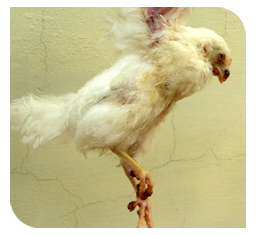
Marek's Disease (MD)
Nature of disease
 |
 |
 |
12-24 weeks of age chicken susceptible to Marek's disease |
Contagious viral disease of poultry |
Gallid herpes virus |
- It is highly contagious viral disease of poultry characterized by enlargement of nerves and internal organs.
- It has great economic significance.
Causes
 |
 |
 |
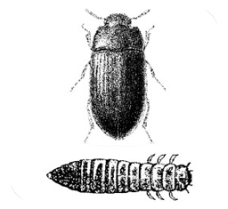
Beetles |
Droppings |
Herpes virus |
- Herpes virus causes this disease.
- Virus can survive at 370C for 24 hrs and in poultry house, droppings, litter, dust, feather follicles and dander for several weeks or months.
- Virus can be inactivated by common chemical disinfectants within 10 minutes of treatment as well as with high humidity.
- The virus matures into a fully infective, envelope form in the epithelium of the feather follicle, from which it is released into the environment.
- It may survive for months in poultry house litter or dust. Dust or dander from infected chickens is particularly effective in transmission.
- Transmission occurs by direct and indirect contacts by air- borne route (aerosols) and inhalation of infective materials mainly dust containing epithelial cells of feather follicles and dander.
- Once the virus is introduced into a chicken flock, regardless of vaccination status, infection spreads quickly from bird to bird.
- Infected chickens continue to be carriers for long periods and act as sources of infectious virus. Shedding of infectious virus can be reduced, but not prevented, by prior vaccination.
- Infective materials can also be transmitted through fomites, personnel and beetles.
- Virus is not transmitted through eggs.
- The incidence of Marek’s disease is quite variable in commercial flocks and depends on strain and dose of virus, age at exposure, maternal antibody, host gender and genetics, other concurrent diseases, and several environmental factors including stress.
Clinical symptoms
 |
.JPG) |
 |
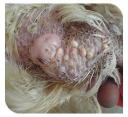
| Enlarged feather folliclees |
Leg paralysis |
Lymphoid tumours in internal organs |
- Disease appears in two distinct forms, viz. acute and classical form.
- Lameness or paralysis around 12 weeks of age may be indicative of this disease.
- Acute form is manifested with sudden deaths due to formation of lymphomas in the visceral organs.
- Birds may show depression, anaemia, anorexia, emaciation and weight loss, diarrhoea, etc.
- Younger birds from 4 weeks onwards (mostly 6 to 10 weeks) generally suffer from this form and mortality may go as high as 60%.
- Classical form involves lesions in the nerves of birds above 12 weeks with 10-30% mortality occurring over a long period.
- Commonly, there is in coordination, lameness, partial or complete paralysis of wings and legs with birds unable to stand.
- A transient paralysis syndrome (unilateral leg paresis) has been associated with Marek’s disease, causing a characteristic posture of one leg held forward and the other held backward as lesions progress.
- Stretching of legs and wings due to paralysis in either direction will give common appearance of split leg in the disease.
- Twisting of neck (torticollis) results from involvement of cervical nerves and paralysis and dilatation of crop results from involvement of vagus nerves.
- Blindness may also occur due to unilateral or bilateral ocular involvement.
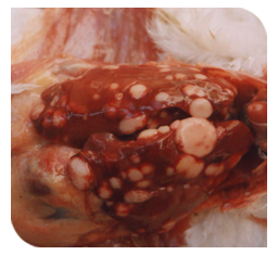
Gross lesions
- Diffuse or nodular lymphoid tumors may be seen in various organs, particularly the liver, spleen, gonads, heart, lung, kidney, muscle, and proventriculus.
- Enlarged nerves are one of the most consistent gross lesions in affected birds.
- Enlarged feather follicles commonly termed skin leukosis may be noted.
Prevention and control
|
|
|
Biosecurity |
Sanitation |
Vaccination |
- Vaccination is the one of the most exploited and economical ways of Marek’s Disease control and solid immunity develops after 7 days of vaccination.
- Recommended vaccination schedule for layers:
Age in days |
Name of vaccine |
Route |
0 |
MD (Bivalent) |
S/C |
7-10 |
MD (Bivalent) |
S/C |
- Now days, in ovo vaccination (in 18 days old embryo) are done with automated technology in many developed countries.
- Procurement of stocks from Marek’s Disease resistant sources followed by regular testing for Marek’s Disease.
- All-in-and-all-out policy of rearing the stocks minimizes the chances of disease in vaccinated flocks by breaking the infection cycle with disinfection.
- Strict biosecurity along with adequate hygiene and sanitation in addition to vaccination are essential for prevention of the disease.
- After outbreaks, premises should be disinfected with 5% formalin and kept without stocks for at least one month.
TOP
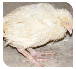
Infectious Bursal Disease (IBD)
Nature of disease
|
|
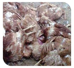
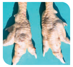
80-90% of mortality |
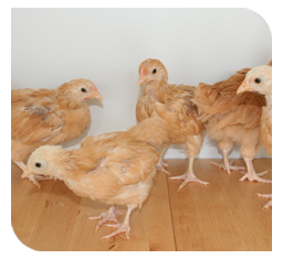
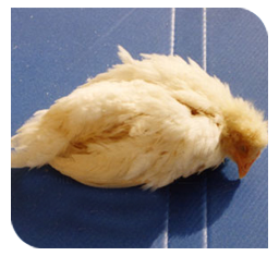
Young chicks upto 0-6 weeks are more susceptible |
- This is acute and highly contagious infection of chickens.
- It is otherwise called as Gumboro disease or Infectious Bursitis or Avian nephrosis.
- Young chicks upto 0-6 weeks are more susceptible.
- Morbidity is 100% and mortality is 80-90%.
- B- Lymphocytes are the primary target cells. It primarily affects the bursa of fabricius, an important organ responsible for immunity.
- Incubation period is short and clinical signs observed in 2-3 days following infection.
- Economically significant, because heavy mortality in 3 – 6 wks old chickens and older and severe prolonged immunosuppression of chickens infected at an early age.
- This disease breakdowns the immunity, leading to the outbreak of other diseases.
- Immunosuppression leads to vaccination failures, Escherichia coli infection, and Gangrenous dermatitis and Inclusion Body hepatitis – anaemia syndrome.
Causes
 |
 |
 |
Birna virus |
Egg trays, vehicles used in the transport of birds and eggs |
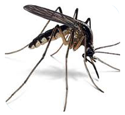
Mosquito |
- The Birna virus belongs to the family Birna viridae and genus Avibirna virus.
- This virus is highly contagious and persistent in the environment of poultry houses.
- Affected birds excrete the virus in faeces for 10-14 days.
- Virus survives upto 120 days in poultry sheds.
- Water, feed, droppings from infected birds are viable for 52 days in the poultry houses.
- Hardy nature of this virus survives heat, cleaning and disinfectant procedures.
- Survives in the environment between outbreaks.
- Meal worm, Aedes vexan (Mosquito) and litter mites appear to act as carriers and remains infective for up to 8 weeks.
- Egg trays, vehicles used in the transport of birds, eggs and personal handling of birds in sheds and elsewhere are very important source of carriers of infection.
- Role of mechanical vectors (Human, wild birds, insects).
- No vertical transmission and carriers. (Disease is not transmitted through eggs).
- Older birds (due to Bursal regression) are more resistant to infection.
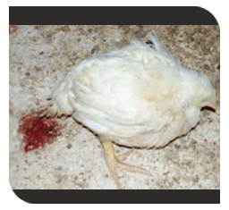
Clinical symptoms
 |
 |
 |
Closed eyes and death |
Ruffled feathers |
Watery and whitish diarrhoea |
- Self vent pecking.
- Anorexia
- Depression and trembling
- Watery and whitish diarrhoea
- Soiled vents
- Ruffled feathers
- Reluctant to move
- Closed eyes and death.
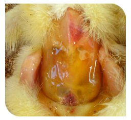
Gross lesions
|
|
|
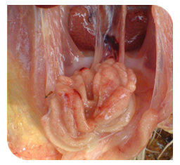
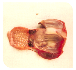
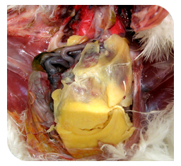
Bursal haemorrhages and enlargement |
Haemorrhages in proventriculus & Gizzard junction |
Musle haemorrhages |
- Dehydration of carcass.
- Petechial / paint brush haemorrhages on the leg, thigh and pectoral muscles.
- Hemorrhage in the Proventriculus and Gizzard junction.
- Enlargement of bursa fabricius to almost double its normal size.
- Haemorrhage on the internal and serosal surfaces of the bursa fabricius.
- Intestine with excess mucus.
Prevention and control
 |
 |
Disposables - Deep burial with slaked lime |
Toxin free feed |
- Primary vaccination with mild or intermediate strain at 2 weeks of age.
- Booster vaccination with intermediate strain (live) after 3 weeks of age.
- Recommended vaccination schedule for layer chicks:
Age in days |
Name of the vaccine |
Route |
12-14 |
IBD Live ( Primary) |
I/O |
22-24 |
IBD Live (Booster) |
I/O |
- Vaccination of breeder stock and seromonitoring by hatcheries to ensure adequate levels of maternal antibodies in the chicks.
- To obtain high levels of MDA in progeny, parent stocks are vaccinated between 4 and 10 weeks of age with live vaccine and again at approximately 16 weeks with inactivated oil-adjuvant vaccine.
- Include immuno-stimulants like Vitamin E in the feed.
- Give toxin-free feed.
- Disposal of litter, dead birds, used gunny bags, curtains and other disposables by incineration or deep burial with slaked lime.
- Restricting vehicular movements with crates, egg trays and culled birds.
- Treating feeders and waterers with 5% formalin.
- Fumigating new poultry sheds with formalin fumes.
- Restricting personnel to their sheds for work.
TOP
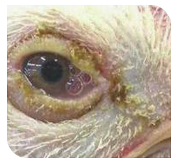
Infectious Coryza
Nature of disease
 |
 |
 |
Discharge from the eyes |
Discharge from the nostrils |
Swelling of the face |
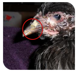
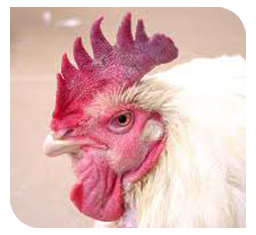
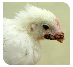
- Infectious coryza is an acute, highly contagious, bacterial disease of the upper respiratory tract of chickens.
- A chronic respiratory disease can develop when complicated by other pathogens.
- Characterized by swelling of the face (facial oedema), and discharge from the eyes and nostrils.
Causes
 |
 |
 |
Drinking water contaminated by discharge |
Homophiles paragallinarum |
Older bird suffers more |
- This disease is caused by a bacteria, Homophiles paragallinarum
- Older bird suffers more severely.
- Clinically affected and carrier birds act as a main source for disease.
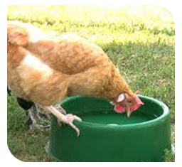
- It can be transmitted by drinking water contaminated by nasal discharge as well as by airborne means over a short distance.
- Lateral transmission occurs readily by direct contact.
- Factors that predispose to more severe and prolonged disease (chronic respiratory disease) include intercurrent infections with microorganisms such as infectious bronchitis virus, Laryngotracheitis virus, Mycoplasma gallisepticum, Escherichia coli or Pasteurella spp. and unfavorable environmental conditions.
- Economic losses are due to marked reduction in egg production (10-40%) in layer.
Clinical symptoms
 |
 |
 |
01 Swelling and cyanosis of eyelids and face |
02 Swelling and cyanosis of eyelids and face |
03 Swelling and cyanosis of eyelids and face |
- The disease in flocks on deep litter management is characterized by rapid spread, high morbidity and low mortality.
- First typical symptoms include sneezing, mucus –like discharge from the opening of the nose, eyes, and swelling on the face(facial oedema)
- In severe case conjunctivitis with closed eyes, swollen wattles, and difficulty in breathing can be seen.
- Feed and water consumption is usually decreased resulting in a drop in egg production.
Gross lesions
 |
Infraorbital sinus showing consolidated caseous exudate |
- Catarrhal to fibrino-purulent inflammation of the nasal passages and infraorbital sinus and conjunctivae.
- As the disease becomes chronic or other pathogens become involved, the sinus exudates may become consolidated and turn yellowish.
- Subcutaneous edema of the face and wattles is prominent.
- The upper trachea may be involved, but the lungs and air sacs are only affected in chronic complicated cases
Prevention and control
- Prevention of infection into the farm is the best control, which could be achieved by hygiene, sanitation, strict bio security and procurement of birds from disease free sources.
- Since recovered birds are reservoir of infection, such birds should be removed and culled from the flock.
- All – in – all- out rearing system is required for eradication of disease.
- Vaccination using inactivated whole culture of organisms containing an adjuvant can protect chickens against the disease.
- In endemic areas, two doses of vaccine, each of which must consist of at least 108 colony-forming units are advocated, given subcutaneously, the first at about 16 weeks of age. Another one is given at 20 weeks of age.
- After cleaning, disinfection and resting of the building for at least 1 week, new birds may be introduced.
- Only day-old chickens or older birds which are known to be free from H. Paragallinarum should be used for the restocking.
TOP
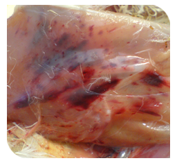
Infectious Bronchitis (IB)
Nature of disease
 |
 |
 |
E.coli increases the severity of disease |
Mycoplasma - increases the severity of disease |
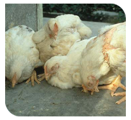
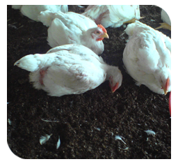
Under 6 weeks age of chicks are more susceptible |
- This is a highly infectious and contagious respiratory disease of chicks. Also affect the oviduct, and some strains have a tendency for the kidneys.
- Disease can occur at any stage, but young chicks, especially under 6 weeks of age are more susceptible.
- Great economic importance due to its adverse effect on egg production and egg quality in layers, and on production in broilers.
- Other pathogen such as Mycoplasma or E.coli increases the severity and duration of the disease.
Causes
 |
 |
 |
Contaminated feed |
Egg shells of infected birds |
Infectious bronchitis virus |
- The virus belongs to corona group of virus.
- Virus is fragile in nature and gets destroyed by common physical and chemical agents.
- Infection is by inhalation of droplets, through ingestion of feed and water contaminated with virus and by contact with infected birds, contaminated movable equipments, clothing and personnel.
- Virus is present in respiratory discharges, faeces and eggshells of infected birds.
- This virus survives well outside the body during winter therefore, disease incidence and spread is more during winter than other seasons, though disease can occur in any season.
- Spreads very rapidly in the flock.
- Bird to bird by direct transmission.
- Transmission through eggs.
- Fomites also can transmit the disease.
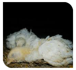
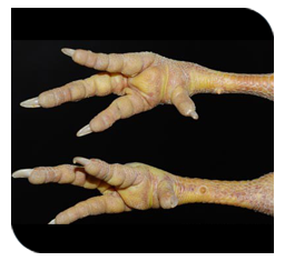
Clinical symptoms
 |
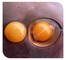 |
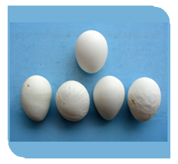
Misshapen egg with ridges |
Watery albumin of egg |
- In young chicks upto 6 weeks of age, respiratory signs like sneezing, coughing, gasping, tracheal rales, lachrymation and nasal discharge are more common.
- The chicks will huddle under the hover.
- There may be swelling of sinuses and face.
- In chicks, mortality may as high as 25%-60% and the course of disease is 1-2 weeks.
- Respiratory noises can be heard more distinctly during night when chicks are normally quite.
- In growers and adult birds, signs of less intensity are seen with less occurrence of nasal discharge and mortality is negligible.
- In laying birds, egg production declines (5-50%) rapidly.
- Damage to functional oviduct in adults (most common).
- Egg abnormalities (Production of misshapen, thin or soft –shelled, rough, smaller, corrugations and leathery eggs).
- The egg quality is poor with thin or watery white albumin.
- In uraemic form, birds exhibit depression, ruffled feathers, wet droppings, increased water intake and increased mortality (0.5-1% per week) due to urolithiasis (Kidney stones).
Gross lesions
- Serous, catarrhal, or caseous exudates in the trachea and bronchi lumen, generally without haemorrhages.
- Plugs of yellow caseous material obstructing the bronchi and lower parts of trachea of chicks that dies.
- Fluid yolk material may be found in the abdominal.
- Abnormal ovary having the misshapen follicle.
- Middle third of the oviduct may appear atrophied and ova ruptured into abdominal cavity.
- Swollen, pale kidneys and deposits of urates in kidney, ureters and throughout the body.
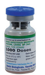
Prevention and control
- Strict hygienic management procedures and vaccination can prevent the disease.
- Recommended vaccination schedule for layers
Age in days |
Name of the vaccine |
Route |
5-7 |
IB Live |
I/O |
28-30 |
IB Live |
I/O |
80 |
IB Live |
D/W |
112-114 |
IB Killed |
S/C |
280 |
IB Killed |
S/C |
TOP
Gout
Nature of disease
|
|
|
Death - due to kidney failure |
Deposition of urates on joints |
Laying hens fed high level of calcium |
- Gout is a not disease condition, but a clinical sings of severe kidney dysfunction.
- Characterized by presence of high level of uric acid in the blood.
- Deposition of urates on the surface of various internal organs or joints (especially hock joint).
- Death is due to kidney failure.
- It is a main problem of laying hens fed high level of calcium.
- Two distinct forms are there visceral gout and articular gout.
Causes
 |
|
 |
Dehydration |
Infectious bronchitis virus |
Treatment with sodium bicarbonate |
- Kidney dysfunction leads to hyperuricaemia.
- Dehydration.
- Excessive dietary calcium or calcium: phosphorus imbalance.
- Vitamin A deficiency.
- Increased intake of protein.
- Intake of excessive amount of salt.
- Infection with infectious bronchitis virus in young chicken.
- Urolithiasis and mycotoxins.
- An electrolyte excess or deficiency.
- Prolonged treatment with sodium bicarbonate.
Clinical symptoms
 |
 |
Deposition of urate salts as a white chalky coating on organs |
Deposition of urate salts as a white chalky coating on organs |
 |
Affected leg joints |
- In articular gout joints are much swollen, with deposition of masses of chalk-like material. Usually wing and leg joints are affected.
- Affected bird cannot move and so die of starvation.
- Articular gout occurs mostly in male birds but visceral gout occurs both in male and female.
Gross lesions
- In articular gout tissue surrounding the joints is white due to urate deposition.
- In Visceral gout kidneys are swollen and congested and greyish white in color. Apart from kidney, chalk-like crystals are deposited on the serous membranes of various internal organs like mesentery, peritoneum, heart, proventriculus and lungs.
- Urate deposition appears as a white chalky coating on organs.
Prevention and control
|
|
Increase maize |
Water containing electrolytes |
- Avoid feeding of high level of calcium in advance of sexual maturity.
- Reduce high level of protein.
- Increase maize, and formulate the feed.
- Give plenty of water containing electrolytes
TOP
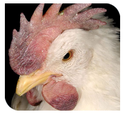
Fowl pox
Nature of disease
 |
 |
Disease affects birds of all ages |
poor weight gain |
- Fowl pox is a common, slow spreading, widely prevalent, contagious viral disease of poultry.
- This is characterized by wart like growth over the non-feathered parts of skin and mucosa of the upper respiratory and digestive systems.
- Disease affects birds of all ages.
- Economically very important as it can cause poor weight gain, drop in egg production in layers and mortality.
- It has no zoonotic importance.
Causes
 |
 |
 |
Avipoxvirus |
Overcrowding |
Through vaccination virus transfer from one to another |
- Disease is caused by an avipoxvirus, which is relatively resistant to common disinfecting procedures and can survive in dried scab for years.
- Virus does not penetrate intact skin. Some break in the skin is required for the virus to enter the epithelial cells, replicate and cause disease.
- Disease spreads mechanically through direct contact, where contaminated materials soil the abraded/lacerated skin, by mosquitoes and other biting insects.
- In a contaminated environment, aerosols (droplets) generated from feather follicles and dried scabs carry the virus and spread the disease.
- It can be transmitted by the respiratory tract.
- During vaccination, the individuals may transfer the virus from affected to healthy birds.
- Deposition of virus in eyes, through lachrymal duct it goes to larynx and causes upper respiratory tract infection.
- Disease is frequently observed during rainy and winter seasons with persisting overcrowding and unhygienic conditions.
- Disease remains a problem for a long time, in farms where multiple age birds are maintained, even after preventive vaccinations.
Clinical symptoms
 |
 |
 |
Wart like gowth in the non-feathered parts of the body |
Wart like gowth in the non-feathered parts of the body |
Wart like gowth in the non-feathered parts of the body |
- This disease is manifested clinically in one of the two forms, cutaneous or diphtheritic or both.
- Poor weight gain and drop in egg production in layers is always significant.
- In cutaneous form (dry pox), lesions in the form of scab appears on the comb, wattles, eyelids, external nares, corner of the beak and other non-feathered parts of the body.
- Lesions on eye may affect the bird’s vision or close both eyes affecting the ability of the bird to reach feed and water leading to starvation and deaths.
- Papule —> vesicles —> pustules —> crust/scab —> scar formation.
- In cutaneous form, flock mortality is usually low rarely exceeding 25%.
- In diphtheritic form (wet pox or fowl diphtheria), initially small nodules are formed on the mucous membrane of mouth, oesophagus and trachea.
- Later on, these nodules become yellow cheesy in nature, thereby, forming a diphtheritic membrane on these organs causing obstruction, interference with feeding and difficulty in breathing with mortality up to 50%.
Gross lesions
 |
fowl pox gross |
- Lesions initially start as nodular area with blanched appearance (papule)
- It becomes enlarged and yellowish (pustules) terminating into thick, dark scab.
Prevention and control
 |
 |
Disinfection of premises |
Sanitation |
- Two types of vaccines (pigeon pox and fowl pox vaccines) can be used for vaccination.
- Pigeon pox vaccine is less pathogenic and can be used on chickens at any stage by wing web method and it produces immunity for 6 months therefore revaccination is required.
- Fowl pox vaccine produces solid immunity, usually carried out at 6-8 weeks of age by intramuscular route or wing web method.
- Successful or effective vaccination can be judged by takes, where examination of vaccinated birds after 7-10 days of vaccination, show swelling or a scab at the site of puncture or vaccine application.
- Absence of takes indicates poor potency of the vaccine, presence of maternal antibodies and improper vaccination.
- In such cases, revaccination with new batch/ lot of vaccine should be done.
- During disease outbreak, affected birds if less than 30% should be segregated immediately and the remaining birds must be vaccinated at earliest possible.
- Standard sanitation and strict biosecurity measures can control the fowl pox.
- Disinfection of premises with sodium hydroxide (1:500), cresol (1:400) and phenol (3%) proved beneficial in control of fowl pox.
TOP
Colibacillosis
Nature of disease
 |
 |
 |
Avian pathogenic escherichia coli. |
Coligranuloma |
Mushy chick disease |
- Colibacillosis is a localized or systemic infection caused by avian pathogenic Escherichia coli bacteria characterized by septicemia, drop in production and mortality.
- Colisepticaemia, egg peritonitis, yolk sac infection(“mushy chick disease” and “omphalitis’), and coligranuloma (Hjarre’s disease) are the well recognized results of E.coli infection. These conditions are collectively grouped under the heading “Colibacillosis”.
Causes
 |
 |
 |
Birds consuming beetles |
Birds in poor environmental condition |
Contaminated eggs |
- Escherichia coli is a normal inhabitant of the digestive tract and wild birds, which are source of infection for poultry flocks and spread is by direct or indirect contacts.
- Organisms are susceptible to common physical and chemical disinfectants. They can survive in freezing and can remain viable for long period at low temperatures.
- In litter, high ammonia concentration can inactivate rapidly and survive at 370C for 1-2 days.
- Trachea, caeca and oviduct of recovered birds can harbor E.coli for several weeks.
- Organisms are not transmitted through eggs.
- Beetles can transmit the bacteria and birds consuming these beetles get the infection.
- Large numbers of E. coli are maintained in the poultry house environment through fecal contamination.
- Initial exposure to pathogenic E. coli may occur in the hatchery from infected or contaminated eggs, but systemic infection usually requires predisposing environmental factors or infectious causes.
- Mycoplasmosis, infectious bronchitis, Newcastle disease, hemorrhagic enteritis, and turkey bordetellosis precede colibacillosis.
- Poor air quality and other environmental stresses may also predispose to E coli infections.
- A hatching environment that is not sufficiently humid is often associated with a high incidence of yolk sac infection. E.coli multiplies rapidly in the intestines of newly hatched chicks and infection spreads rapidly from chick to chick in the hatchery and brooders.
Clinical symptoms
 |
 |
 |
Depression and disinclination to move |
Mortality in a batch of chicks - first week after hatching |
Swollen Head Syndrome |
- Affected birds will have respiratory symptoms, depression, loss of appetite and disinclination to move.
- Soiling of vent with pasty diarrhoeic faeces will be seen.
- In Coliform omphalitis/yolk sac infection causes mortality in a batch of chicks in the first week of life after hatching. Affected chicks exhibit depression, sleepiness with tendency to huddle together around heat sources with distended abdomen and swelling of naval.
- Swollen Head Syndrome (SHS) causes swelling of head.
Gross lesions
 |
 |
 |
Cassiated egg mass inside the oviduct |
Egg peritonitis |
Inflammed unabsorbed yolk sac with abnormal colour |
- Birds with Colisepticaemia develop airsacculitis, perihepatitis and pericarditis with cloudiness of pericardial sac and light coloured fibrinous exudates.
- In Coliform omphalitis/yolk sac infection, unabsorbed yolk will have abnormal volume, colour, consistency and smell.
- Salphingitis (inflammation of oviduct) causes peritonitis, abdominal cavity containing abnormal egg or yolk mass, impaction of oviduct, distortion of ovaries and produce foul smelling pus like material.
- Coligranuloma (Hjarre’s disease) causes millet sized multiple projected granulomas are found on the surface of liver, caeca and mesentery.
- Air sac disease (Chronic Respiratory Disease) causes thickened air sac and will have caseous exudates.
- A whole or partly formed egg may be seen in the abdominal cavity known as impaction of oviduct which was firm and adhered to peritoneum and visceral organs.
Prevention and control
 |
Biosecurity |
- Treatment strategies include attempts to control predisposing infections or environmental factors and early use of antibacterials indicated by susceptibility tests.
- Maintain adequate hygiene and bio security to minimize transmission through faecal contamination of eggs.
- Infected excreta and litter should be disposed properly to avoid contamination of natural water sources and spread in the farm.
- Avoid stress and overcrowding in the flocks which favors outbreaks.
- Birds should be procured from sources tested free of ND, IB, and Mycoplasma.
- Diet with protein, selenium, and vitamin E may be favorable in control of colibacillosis.
- Chlorination of drinking water inactivates the bacteria.
TOP
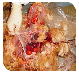
Coccidiosis
Nature of disease
 |
Heavy mortality in broilers |
- Coccidiosis is one of the most important protozoan diseases of poultry.
- Outbreaks are common between 3- 6 weeks of age.
- Inflict heavy mortality in broilers and also in growers raised on deep litter.
- One of the biggest causes of economic losses to poultry.
Causes
 |
 |
rodent |
Seven species of genus Eimeria |
- It is caused by seven species of genus Eimeria.
- There is no cross immunity (cross-protection) among the seven species of Eimeria.
- Of the seven disease-producing species, E.tenella, E.necatrixand E.brunettiare the most harmful and cause high morbidity and high mortality.
- E. tenellaaffects caeca and E. necatrixmiddle portion and E. brunettilower portion of the small intestine.
- E. maximaand E. acervulinaare moderately harmful. E. maximaaffects middle portion of the small intestine, and E.acervulinamainly upper portion, that is, duodenum.
- Ingestion of the infective form of oocysts (sporulated oocysts) is the only natural method of spread.
- Ingestion of feed and water contaminated with sporulated oocysts causes the infection.
- Infected chickens may shed oocysts in the faeces for several days or weeks.
- Oocysts can be spread mechanically by movement of people, equipment and foot wear between farms.
- It can also spread through cockroaches, rodents, pets and wild birds.
Clinical symptoms
 |
|
 |
Bloody diarrhoea |
Dehydration |
Drooping wings |
- Affected birds appear dehydrated with drooping wings and ruffled feathers.
- They may huddle together and severe watery or bloody diarrhoea.
- Droppings of affected birds usually contain blood, fluid, and mucus.
- Emaciated and anaemic appearance.
- High mortality and most occurs between 5 and 6 days following infection.
- Causes poor weight gain and
- Egg production may be reduced in laying birds.
Gross lesions
 |
 |
 |
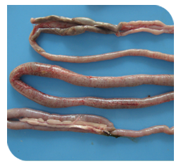
Distended small intestine filled with fluids & clotted blood |
Distended small intestine filled with fluids & clotted blood |
Distended small intestine filled with fluids & clotted blood |
- Caeca enlarged with clotted blood.
- Middle portion of the small intestine is distended to twice its normal size.
- Intestinal lumen filled with blood.
- Lining of the small intestine covered with tiny hemorrhages.
- Intestinal mucosa swollen and thickened.
Prevention and control
 |
 |
 |
Coccidiosis Vaccine |
Use lime powder to dry the litters |
Use slphonamide as feed additive |
- Coccidiosis is far more easily prevented than treated.
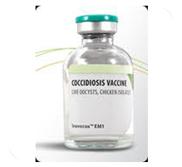
- Control depends mainly on drugs, although an effective vaccine is now available for breeder or layer replacements.
- Two types of vaccines have been used to obtain immunity (protection) against coccidiosis.
- Birds are vaccinated through drinking water between the age of 5 and 9 days.
- The use of one anticoccidial in the starter and another in the grower feed is called a 'shuttle programme'.
- The use of shuttle programme has been found to reduce drug resistance.
- Hygiene and biosecurity along with measures to prevent contamination of feed and water with droppings can prevent the occurrence of infection.
- Dry litter and raking of litter at regular intervals prevent sporulation of oocysts, thereby minimizes chances of infection.
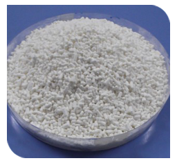
- During rainy season lime powder can be used to dry the litter and to render the oocysts ineffective.
- Use of anticoccidial in feed as feed additives (e.g.; lonophores, sulphonamides, and quinolones) can avoid acute outbreak.
- Rotation of anticoccidial every 4-6 months are helpful in maintaining their efficacy and restricting the development of resistance.
- In disease condition, medication must be given in consultation with qualified veterinarian to avoid complications.
TOP
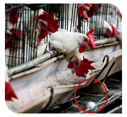
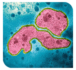
Avian Influenza (Bird flu)
Nature of disease
 |
|
Drop in egg production |
Heavy mortality in broilers |
- Coccidiosis is one of the most important protozoan diseases of poultry.
- Outbreaks are common between 3- 6 weeks of age.
- Inflict heavy mortality in broilers and also in growers raised on deep litter.
- One of the biggest causes of economic losses to poultry.
Causes
 |
 |
 |
Disease is not transmitted through eggs |
Influenza virus type A. |
Wild birds transmit disease from one place to another |
- This is caused by Influenza virus type A.
- This virus present in body secretions and faeces are very resistant to common physical (heat, sunlight, dryness, etc.) and chemical (formaldehyde, sodium hypochlorite, etc.) factors and can survive for many days at low environment temperature.
- This disease is transmitted mainly by direct contact between birds and by air (aerosols) and fomites (equipments, shoes, clothing, vehicles, etc.).
- Virus is excreted in all secretions and faeces of infected bird, which contaminate the environment and all poultry appliances/ equipments.
- Wild birds are also known to transmit disease from one place to other.
- Disease is not transmitted through eggs.
Clinical symptoms
 |
 |
 |
Haemorrhages in thigh muscles |
Swollen and cyanotic face, wattle and comb |
Swollen and subcutaneous haemorrhages in feet and shank |
- Mildly pathogenic Avian Influenza pathotypes affect respiratory, digestive, urinary and reproductive organs of poultry and all symptoms associated with these systems are observed.
- Affected birds will have coughing, sneezing, rales, anorexia, depression, drop in egg production, Softened egg shell and diarrhoea.
- Swollen and cyanotic wattle and comb.
- Sero-mucous nasal discharges and hypersalivation.
- Subcutaneous feet petechiael haemorrhages.
- Highly pathogenic Avian Influenza pathotypes cause damage to almost all organs and birds may die without showing any clinical sign.
- Birds will show decreased activity and sounds thereby poultry houses will appear quiet.
- Involvement of nervous system is reflected with tremors of head and neck, inability to stand, torticollis, stiffening of body and abnormal positioning of head.
- Very high mortality rate (almost 100%).
Gross lesions
- Subcutaneous tissues of feet shows petechial haemorrhages.
- Haemorrhages in thigh and chest muscles.
- Hyperemia of trachea.
- Haemorrhages in internal organs.
Prevention and control
- Strict biosecurity measures can prevent the introduction of disease through infected birds into the farm.
- Vaccines can prevent clinical signs and death. Furthermore, viral replication and shedding from the respiratory and GI tracts may be reduced in vaccinated birds.
- Proper disposal of the infected birds and their secretions and excretions followed by proper disinfection of poultry houses and equipment with sodium hypochlorite (5.25%), sodium hydroxide (2%), sodium carbonate (4%) and phenolic compounds.
- Complete destruction of eggs, feed ingredients, litter materials, affected birds in deep pit with addition of lime.
- Restriction of movement of poultry equipment and people within the farm during outbreak.
- The contact between wild birds and domestic birds should be avoided, as former are reservoirs of infection.
- Burning, burying or composting can destroy viruses in litter and manure.
- Inactivated influenza virus vaccines can be effective in reducing the mortality and production losses. However, immunity produced is subtype-specific and is not practical to use preventive vaccination for all subtypes. Vaccine can be useful where virus subtype is identified.
- Suspected outbreaks should be reported to appropriate regulatory authorities.
TOP
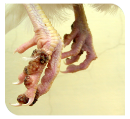
Ascariasis
Nature of disease
 |
 |
Anaemia and weight loss |
Young birds up to 3 months of age are more susceptible |
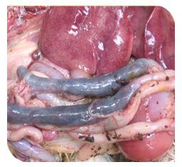
- This is a worm infestation of chickens.
- Young birds up to 3 months of age are more susceptible.
- It has significant effect on the production of birds.
- It causes diarrhoea, anaemia, weight loss, increased mortality and reduced egg production.
Causes
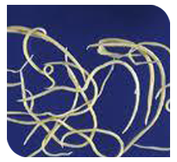 |
 |
 |
Ascaridia galli |
Malnourished bird |
Reusing of litter materials |
- Ascaridia galli is the species found in intestines of chickens.
- Ascaridia are the largest roundworm of birds.
- Parasites usually affect malnourished flocks.
- Serious infection occurs if the litter is reused in the case of broilers.
- Dietary deficiencies such as vitamin A, B and B12, various minerals and proteins leads to heavy infection.
- A.galli eggs are ingested by grass hoppers or earth worms, hatch, and are infective to chickens.
- Under optimum conditions of temperature and moisture, eggs in the droppings become infective in 10 – 12 days.
- Eggs are quite resistant to low temperatures.
- Chicken over 3 months are more resistant to infection.
Clinical symptoms
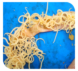 |
 |
 |
Occlusion of thread like worms in intestine |
Anaemic |
Decreased egg production |
- Cause poor bodily condition and weight loss.
- Birds become anaemic and suffer from diarrhoea.
- In heavy affection, it causes haemorrhagic enteritis.
- Loss of blood and increased mortality.
- Affected birds become unthrifty, markedly emaciated and egg production is decreased
Gross lesions
- Occlusion of thread like worms in the intestine.
- In heavy infection, intestinal obstruction may occur.
- Ascaridia wormsmay found in the hen's egg. Infected eggs can be detected by candling.
Prevention and control
 |
 |
 |
Changing litter material |
Clean feeding troughs |
Clean feeding and water |
- Regular deworming of chicks with piperazine compounds is highly effective.
- Young birds should be separated from older birds.
- Avoid reuse of litter materials.
- Changing of litter can reduce infections.
- Treatment of the soil or litter to kill intermediate hosts is useful.
- Litter may be treated with suitable insecticides.
- Litter materials should always be kept in dry condition.
- Extreme care should be taken to ensure that feed and water are not contaminated.
- Provide clean feeding troughs and drinking water appliances.
- Poultry runs should be well drained.
Vaccination schedule for Broilers
Days |
Vaccine |
Route |
0 day |
Mareks Disease Vaccine
(HVT) |
0.2ml S/C |
5-7 days |
Ranikhet Disease Vaccine- RDVF |
O/N |
10th day |
Infectious Bronchitis vaccine |
O/N |
12-14 days |
Infectious Bursal Disease
Vaccine- Intermediate Georgia |
O/N |
28th day |
Booster RD La Sota |
Water |
Vaccination Schedule for layers
Days |
Vaccine |
Route |
0 day |
Mareks Disease Vaccine (HVT) |
S/C 0.2 ml |
5-7 days |
Ranikhet Disease Vaccine- RDVF |
O/N |
10th day |
Leechi Disease Vaccine (Optional) |
Water |
12-14 days |
Infectious Bursal Disease Vaccine- Pruning Intermediate Georgia |
O/N or water |
18-22 days |
Infectious Bronchitis |
O/N or water |
24-27 days |
IB Vaccine Booster |
Water |
28-30 days |
RD vaccine Booster- La Sota |
Water |
6th Week |
Fowl Pox Vaccine or Infectious Coryza Vaccine (if prevalent in the area) |
S/C |
8th Week |
RD vaccine- RDVK or R2B |
S/C or I/m |
9th Week |
Fowl Pox Vaccine |
Wing web |
12th-13th Week |
IB Booster |
Water |
18th week |
RD Booster- RDVK or R2B |
S/C or I/m |
45th-50th Week |
RD La Sota repeated every once in 2 Months |
Water |
TOP